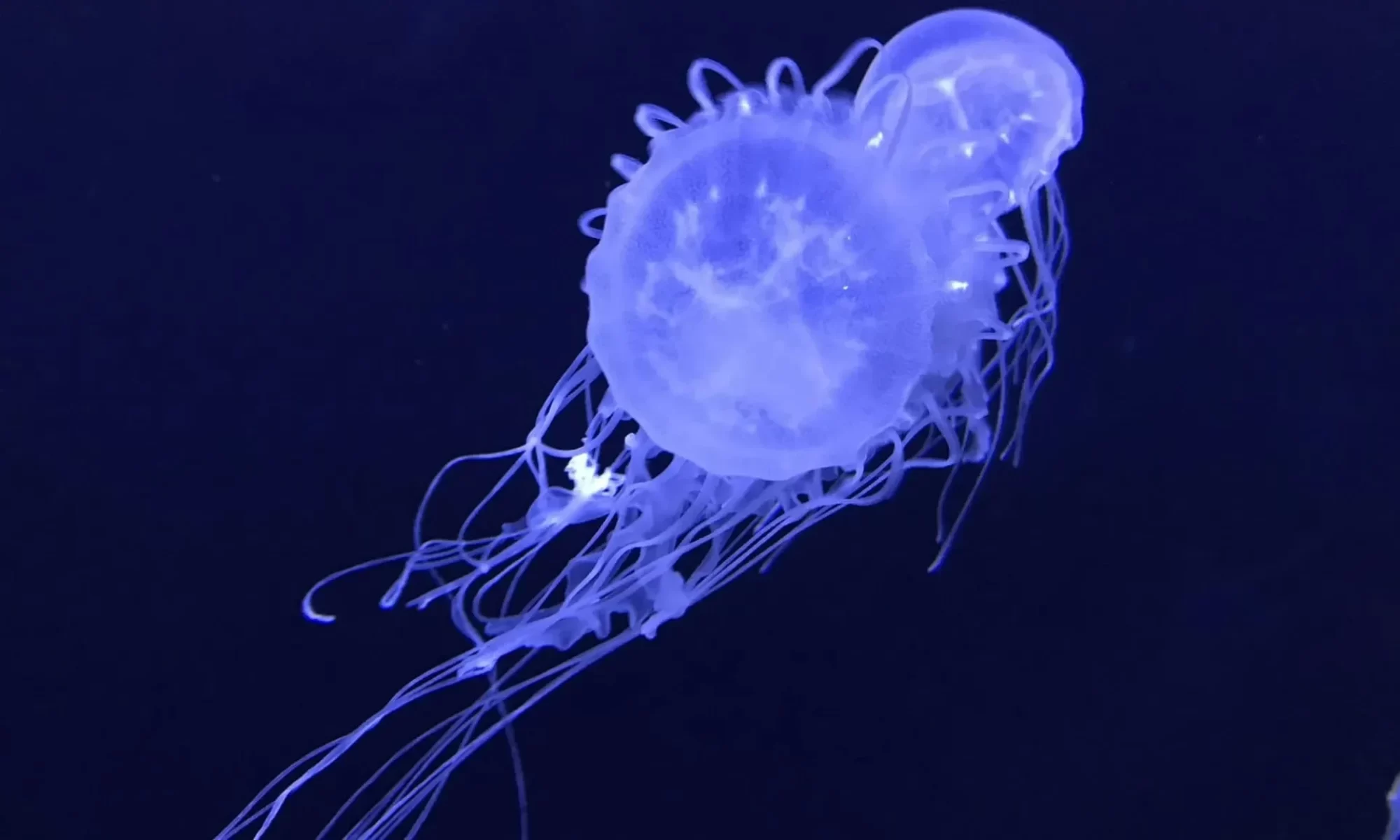
Video showing footage of Cassiopea upside-down jellyfish in Key Largo mangrove forest waters (Florida Keys, USA). Water samples were taken from this collection site during a jellyfish environmental DNA (eDNA) metabarcoding study by Ames et al. 2021, published in Frontiers of Marine Science. Copyright A.C. Morandini (coauthor on the study) “Fieldable Environmental DNA Sequencing to Assess Jellyfish Biodiversity in Nearshore Waters of the Florida Keys, United States”
Tracking Down Ocean Species On the Go Using eDNA

The article highlights the pioneering work of our own Dr. Cheryl Ames and her team in the Florida Keys. They led a crucial study demonstrating the potential of a portable Nanopore sequencer to detect the presence of upside-down jellyfish (Cassiopea xamachana) and other jellyfish species directly in the field! This represents an incredible leap forward, overcoming the challenges of bringing complex DNA analysis from labs into the marine environment.

This technology not only identified Cassiopea jellyfish but also revealed the presence of other species unseen at the time of sampling, such as moon jellyfish and venomous box jellyfish. The potential is vast: from predicting jellyfish sting risks to aiding fisheries management, supporting conservation efforts, and even integrating into autonomous underwater vehicles for comprehensive marine surveys.
This groundbreaking research stems from a foundational scientific paper published in Frontiers in Marine Science, underscoring its impact.
Read the full article on Smithsonian Ocean and dive into the future of species detection:
And for the original scientific paper that underpins this amazing work, access it here:
Fieldable Environmental DNA Sequencing to Assess Jellyfish Biodiversity in Nearshore Waters of the Florida Keys, United States

Recent advances in molecular sequencing technology and the increased availability of fieldable laboratory equipment have provided researchers with the opportunity to conduct real-time or near real-time gene-based biodiversity assessments of aquatic ecosystems. In this study, we developed a workflow and portable kit for fieldable environmental DNA sequencing (FeDS) and tested its efficacy by characterizing the breadth of jellyfish (Medusozoa) taxa in the coastal waters of the Upper and Lower Florida Keys. Environmental DNA was isolated from seawater collection events at eight sites and samples were subjected to medusozoan 16S rRNA gene and metazoan mitochondrial cytochrome oxidase 1 gene profiling via metabarcoding onsite. In total, FeDS yielded 175,326 processed sequence reads providing evidence for 53 medusozoan taxa. Our most salient findings revealed eDNA from: (1) two venomous box jellyfish (Cubozoa) species, including taxa whose stings cause the notorious Irukandji envenomation syndrome; (2) two species of potentially introduced stalked jellyfish (Staurozoa); and (3) a likely cryptic species of upside-down jellyfish (Scyphozoa). Taken together, the results of this study highlight the merits of FeDS in conducting biodiversity surveys of endemic and introduced species, and as a potential tool for assessing envenomation and/or conservation-related threats.
SciShow Explains How Jellyfish Sting Without Touching You!
I Don’t Think You’re Ready for this Jelly!
SciShow’s Olivia Gordon discusses our recent discovery about Upside-Down Jellyfish and how they sting. This jellyfish might look kind of unassuming, but it’s got some surprising long-range weaponry to catch its prey!
Upside-down jellyfish release venom-filled ‘bombs’ in their snot
In a fascinating report by Nicoletta Lanese for Live Science, learn why the water surrounding upside-down jellyfish often stings to the touch. Scientists finally have the answer! Read the full story here
Jellyfish Can Sting You Without Even a Single Touch
These jellyfish can sting without touching you, thanks to ‘mucus grenades’


Imagine encountering a creature that defends itself with unseen projectiles! National Geographic invites you on a journey into the remarkable world of upside-down jellyfish, showcasing Dr. Cheryl Ames’s extraordinary research into their unique defense strategy.
This captivating feature explores how these intriguing invertebrates, Cassiopea spp., can release ‘mucus grenades’ – microscopic, venom-filled structures that pack a potent sting without direct contact. Dr. Cheryl Ames, a marine biologist and associate professor at Tohoku University, provides a fascinating glimpse into the observation of these creatures’ efficiency:
“Then, within 24 hours, the pink cloud will be gone.” (referring to the cloud of zapped brine shrimp after feeding). This observation speaks volumes about the efficacy of their hidden defense system.
National Geographic beautifully illustrates how Dr. Ames’s profound contributions are reshaping our understanding of marine ecosystems and the intricate adaptations within them. It’s truly inspiring to see this deep dive into nature’s secrets featured by such a world-renowned publication, bringing the wonders of the ocean floor to life for millions worldwide.
Upside-down jellyfish release venom-filled ‘bombs’ in their snot


For years, swimmers have felt a perplexing tingling in waters inhabited by upside-down jellyfish, even without direct contact. Now, Live Science delivers the definitive scientific explanation, thanks to the pivotal research co-led by Dr. Cheryl Ames!
The long-standing question of what causes this ‘stinging water sensation’ has finally been addressed. As Dr. Cheryl Ames, an associate professor of applied marine biology at Tohoku University, explains the initial scientific challenge:
“We knew it had to be something in the mucus.”
Her team’s methodical investigation led to the identification of ‘cassiosomes’ – microscopic, venom-filled structures released by the jellyfish – providing the clear answer.
This compelling Live Science article details the journey of scientific inquiry, highlighting the rigorous process that unveiled nature’s hidden mechanisms. We’re proud to see Dr. Ames’s dedication to solving marine mysteries recognized by Live Science, bringing clarity and understanding to a broader audience.
Upside-down Jellyfish Create ‘Stinging Water’ That Kills Prey by Launching Mucus ‘Grenades’
[This study] began when I and other marine biologists were concerned about the source of ‘stinging water’—an irritating sensation that occurred while in the mangrove forest waters studying upside-down jellyfish, and working together with aquarists at major public aquariums,” Cheryl Ames, an author of the study from Tohoku University, Japan, and the Smithsonian’s National Museum of Natural History, told Newsweek.
“There were several theories exchanged by fellow marine biologists, and comments posted online by people after experiencing stinging water during snorkeling or swimming in those areas. We wanted to find out the scientific explanation behind the long-standing stinging water puzzle,” she said.
Cassiosomes are stinging-cell structures in the mucus of the upside-down jellyfish Cassiopea xamachana

Snorkelers in mangrove forest waters inhabited by the upside-down jellyfish Cassiopea xamachana report discomfort due to a sensation known as stinging water, the cause of which is unknown. Using a combination of histology, microscopy, microfluidics, videography, molecular biology, and mass spectrometry-based proteomics, we describe C. xamachana stinging-cell structures that we term cassiosomes. These structures are released within C. xamachana mucus and are capable of killing prey. Cassiosomes consist of an outer epithelial layer mainly composed of nematocytes surrounding a core filled by endosymbiotic dinoflagellates hosted within amoebocytes and presumptive mesoglea. Furthermore, we report cassiosome structures in four additional jellyfish species in the same taxonomic group as C. xamachana (Class Scyphozoa; Order Rhizostomeae), categorized as either motile (ciliated) or nonmotile types. This inaugural study provides a qualitative assessment of the stinging contents of C. xamachana mucus and implicates mucus containing cassiosomes and free intact nematocytes as the cause of stinging water. The discovery of cassiosomes is detailed in this paper
Click here to access the full article.
Click here to see the media impact of the discovery of cassiosomes