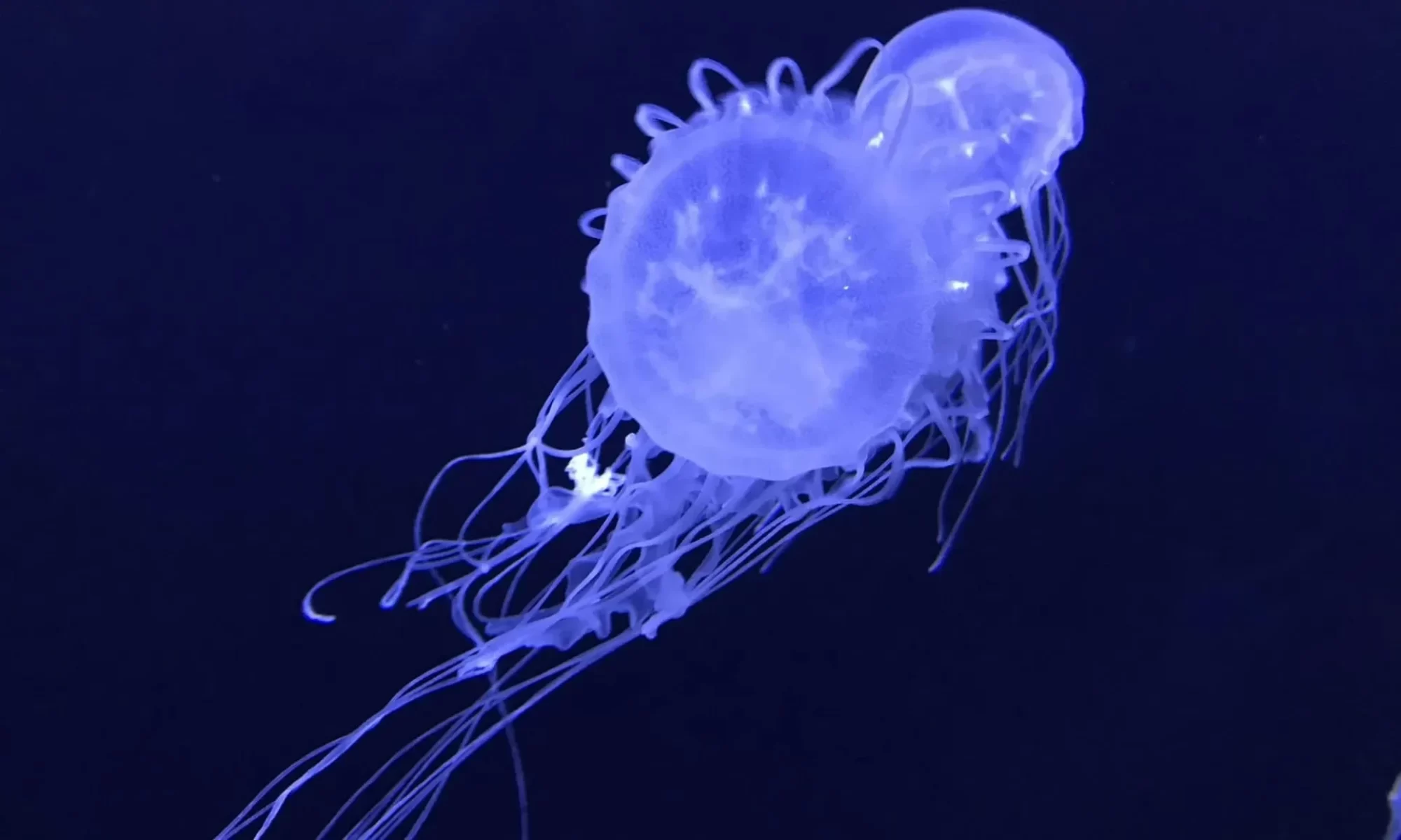
Chironex box jellyfishes (Cnidaria: Cubozoa: Chirodropida) in Singapore: Chironex blakangmati, new species, and range extension of C. indrasaksajiae

New Discovery: A New Species of Deadly Box Jellyfish Uncovered!

Iffah Iesa, Cheryl Lewis Ames, Nicholas Wei Liang Yap & Danwei Huang

Abstract. Two venomous box jellyfish species of the genus Chironex Southcott, 1956 (Cnidaria: Cubozoa: Chirodropida) were collected from Singapore’s coastal waters: Chironex indrasaksajiae Sucharitakul, 2017 and a novel species described herein as Chironex blakangmati, new species.
Chironex indrasaksajiae was collected from both the Johor and Singapore Straits around mainland Singapore. Chironex blakangmati, new species, was collected from Sentosa Island along the Singapore Strait and is the fourth species described in the genus. While Cblakangmati’s volcano-shaped pedalial canal and tentacle number are similar to Cyamaguchii Lewis & Bentlage, 2009, its elongated, sharp-tipped velarial canals and DNA sequences distinguish it from other Chironex species. Comparisons of Chironex blakangmati, new species, with C. yamaguchii and C. fleckeri
reveal novel morphological differences at the terminal end of the perradial lappet along the velarium edge, where C. blakangmati, new species, lacks velarial canals extending from the perradial lappet terminus. Juvenile Chironex yamaguchii specimens were examined and ontogenetic variations of velarial canals are herein reported. Preliminary cnidome analysis reveals eight types of nematocysts observed in C. blakangmati, new species, five types in C. indrasaksajiae, and five types in C. yamaguchii. Molecular phylogenetic reconstruction places C. blakangmati, new species, in a clade distinct from its congeners, as sister group to C. yamaguchii based on 16S rRNA gene analysis but diverging earlier than the clade comprising C. yamaguchii and C. indrasaksajiae based on cytochrome c oxidase subunit I gene analysis for which sequence data are comparatively limited. Understanding the biodiversity and seasonality of venomous cubomedusae will help mitigate the risk they pose to human health and safety during maritime activities.

Read the full article here: Raffles Bulletin of Zoology

From Brasil to Japan

Cheryl Ames presented the ship to the Japanese public.

It was an online tour during which Professor Ames presented the living areas and kitchen, commenting on everything from food options to the work schedule. Then, she showed the lab where her teammates and other international scientists were performing extractions on previously collected samples.

The tour was especially interesting because it showcased the entire ROV support and command structure while the ROV was active at a depth of over 500m. This made it possible to see the “behind-the-scenes” of the live ROV dive happening simultaneously on YouTube.

The tour helped convey the scale of the endeavor and the massive infrastructure required to collect data from the mid-water—from the initial dives to the final extractions.

Stay tuned for more mission updates

Meet our lab team aboard Falkor (too)

Falkor (too) has sailed! On board is a diverse group of scientists and specialists composing the cruise crew, working in a collaborative effort to explore the midwater column along the Brazilian coast.

The Schmidt Ocean Institute organized a profile of the current research crew for the “Designing the Future 3” cruise, which includes members of our lab.

Lab members on board:

If you are interested in learning more about the current cruise, find more information on the “Designing the Future 3” page at the Schmidt Ocean Institute website: https://schmidtocean.org/cruise/designing-the-future-3/

OceanShot: The Cruise – the Lab Team Embarks on the RV Falkor (too)

We’re officially off!

The Lab team — Prof. Cheryl Ames, Chanikarn Yongstar (Nune) and Kei CHloe Tan — is already on board in Salvador, Brazil, prepping for the ambitious OceanShot cruise aboard the RV Falkor (too). While the team is currently settling in and getting the gear ready, the ship is set to depart on 15/04, navigating the coastline toward Fortaleza until May 1st. Throughout the journey, the team will be focusing their research on the mid-water column, utilizing a diverse range of scientific methods to analyze this deep-sea “twilight zone.” The best part? You can join the expedition from home! We invite you to follow the journey and watch live ROV dives through the ship’s website at schmidtocean.org/technology/live-from-rv-falkor/.

Stay tuned for more updates from the Atlantic very soon!

From the beginning of February to the end of April, the IMS Lab has the pleasure of being joined by Mr. Nicolo Ferraris from Wageningen University. During his 3-month exchange program, Nicolo will be assisting on the HABs research project.

Statement from Nicolo:

Hi! I’m Nicolo, an Italian MSc Biology student from Wageningen University & Research in the Netherlands, where I specialized in Molecular Ecology. I just finished my thesis on investigating sex determination in invasive lionfish (Pterois miles), which got me hooked on combining lab work with bioinformatics. I’ve always been really interested in applied molecular biology, especially next-generation sequencing technologies like Nanopore for biodiversity monitoring.

I arrived in Sendai on February 1st and have been loving the experience so far. The people in the lab are great and made me feel right at home since day one. I’ve been helping Sang with organizing samples, joined him for field sampling out in Onagawa (which was amazing!), and have been assisting with eDNA extractions and Planktoscope processing. During the Nanopore training workshop in mid-February, I got hands-on experience with library prep and running the GridION for environmental DNA samples across different locations and gene regions. After the workshop, I noticed the bioinformatics workflow was a bit over-complicated, so I introduced the team to EPI2ME, Oxford Nanopore’s platform, and gave everyone a quick tutorial on how to use it. Since then, I’ve been working through the sequenced samples and generating preliminary taxonomic results for our HAB species and broader plankton communities. Outside of lab work, I’ve been enjoying joking around with the friends I made here, playing basketball and volleyball, indulging in the excellent food, and exploring Sendai!

New Research: A comparative molecular study of rhizostome jellyfishes from Japan reveals variability in Symbiodiniaceae taxon associations and cassiosome production.

Led by Kei Chloe Tan , this study dives deep into the complex world of Rhizostomeae that play massive roles in Asian ecology and industry.

What makes these jellies unique?

Rhizostome jellyfishes are unlike your average medusa. They don’t have long, trailing tentacles neither other structures at the eadg of theyr bell; they have eight highly branched oral arms, along which there are suctorial minimouth orifices.

Our study focused on three main goals:

  • Identity: Using DNA barcoding to accurately identify species in Japanese waters.
  • Partnership: Mapping which types of algae live inside which jellyfish.
  • The Sting: Identifying which species produce cassiosomes—the “mucus grenades” responsible for the mysterious “stinging water” syndrome.

Key Discoveries

  • A New Neighbor: We discovered that Cassiopea (Upside-down jellyfish) found in Kagoshima likely represents a new, undescribed species.
  • Symbiotic Flexibility: We found that these jellyfishes are “plastic” in their partnerships, meaning the same species of jellyfish can host different types of algae (Symbiodinium, Cladocopium, or Durusdinium) depending on the individual.
  • The “Grenade” Evolution: We confirmed that cassiosome production is a unique trait of the suborder Kolpophorae. Interestingly, we found that young jellies might not produce them until they reach a certain developmental stage.

“This research provides a baseline for understanding how jellyfish and algae co-evolve, helping us manage coastal ecosystems and the jellyfish fisheries that are so vital to the region.”Chloe Tan, Lead Author

Lab Work in Action

This paper wasn’t just about sequences; it combined advanced molecular techniques with classic microscopy.

  • Tandem DNA Amplification: Chloe developed a new method to simultaneously sequence the DNA of both the jellyfish host and its tiny algal residents.
  • Microscopy: We captured high-resolution images of the cassiosomes—clusters of stinging cells (nematocysts) with algae at their core—proving exactly how these “contactless stings” work.

Read the Full Paper

Interested in the molecular data or the high-resolution phylogenetic trees? You can find the full study here:

カツオノエボシは1種ではなかった:5種への再分類と新種「ミカヅキノエボシ」の命名 執筆:彩恵 りり

サイエンスライターの彩恵りり氏が、カツオノエボシ属(Physalia属)が5種に再分類された最新の研究について詳しく解説してくださいました。これら新種のうち1種の発見に当研究室が貢献したことを、大変光栄に思います。

[彩恵りり氏による解説記事はこちら]

[当研究室のオリジナル論文はこちら]

Nikkei reported on Mikazuki no Eboshi: 宮城県で新種のクラゲ、伊達政宗にちなんで命名 東北大学

Our lab’s discovery was featured in a Nikkei article. The piece focused on the computer simulation used to estimate the period and travel path of the specimen, as it originated from a different region than the discovery site:

コンピューターを使ったシミュレーション(模擬実験)の結果、神奈川県沖の相模湾などから海流に乗って運ばれた可能性があると分かった。研究成果をまとめた論文は学術誌「フロンティアズ・オブ・マリン・サイエンス」に掲載された。

Find the original material at:

Find the original scientific paper here: